How Prostate Cancer MRIs Makes Biopsies Unnecessary

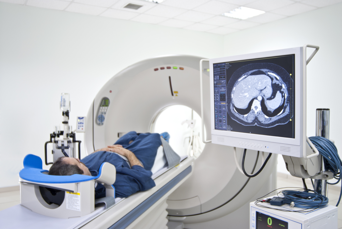
A recent study published in Lancet has shown that multiparametric MRIs are an effective way to screen men with suspected prostate cancer.  The research, conducted a team from the University College London, indicates that prostate cancer MRIs may eliminate the need for a needle biopsy in 25% of men who are suspected of having prostate cancer. Multiparametric MRIs can also measure tumor sizes, which may be useful for detecting aggressive cancers. TRUS biopsies are an invasive procedure that often cause discomfort for patients. Eliminating the need for this procedure in some patients is a goal worth pursuing, because two thirds of biopsies turn out negative for life-threatening cancer.

Today, doctors typically recommend a TRUS biopsy when a patient’s blood test indicates high levels of prostate-specific antigen (PSA). Unfortunately, the procedure often brings side effects such as bleeding, pain, erectile dysfunction and infection. Even if there are no side effects, this type of biopsy requires the insertion of an ultrasound probe into the patient’s rectum, before guiding a needle into the prostate for samples. The procedure takes samples in a random manner, so the results aren’t always accurate. It is always best to avoid this uncomfortable biopsy when possible.

The study analyzed 576 men who were suspected of having prostate cancer. Multiparametric MRI scans detected clinically important cancers in 93% of the men compared to just 48% with TRUS biopsies. Patients with negative scan results ended up having no cancer or a harmless cancer 89% of the time. The results indicate that a TRUS biopsy is unnecessary in about a quarter of men who undergo the scan, while combining this MRI with a TRUS biopsy improves prostate cancer diagnosis. While more research is needed to confirm this approach’s cost-effectiveness, it is a step in the right direction for identifying prostate cancer.
Cancer Death Rate Plummeting in Recent Decades

doctor

The American Cancer Society has published a report showing a dramatic 25% decrease in the cancer death rate since 1991. The ACA has issued the report annually to measure the amount of cancer cases, deaths and survivals on a national level. The mortality rate is expected to continue to drop in the coming years due to widespread lifestyle changes and advances in early detection. Decreases in lung cancer have been an especially large contributor to the lower death rates.

The study found that the cancer rate is likely to be dropping due to a decrease in the number of smokers, especially among men. Cancer deaths among men have fallen 31% since 1990 and 21% among women since 1991, which is largely attributed to decreasing tobacco use. (Men still have a higher rate of cancer deaths than women overall, however.) The Surgeon General’s 1964 report on smoking has changed mindsets about health issues related to smoking.

In the early 90s, there was a large racial disparity for cancer deaths in the US. This issue was especially pronounced for black males, who experienced much higher rates of cancer deaths than the white population. The difference in cancer deaths between black and white men shrunk from 47% in 1990 to 21% in 2014. For women, the difference narrowed as well, from a peak of 20% in 1998 to a low of 13% in 2014. This racial disparity may have decreased due to improved access to healthcare in recent years. The number of uninsured black men and women was cut in half from 2010 to 2015.

Lung, breast, prostate and colorectal cancers saw the highest survival rate improvements. Early detection for these cancers is a key factor in lower mortality, and technological advancements in scanning equipment are sure to have played a role. Detecting cancer is becoming faster, easier and cheaper than ever before. Doctors and patients alike can expect a continuation of the improvements seen in recent years. There may not yet be a 100% survival rate just yet, but the statistics are showing a steady decrease in deaths, which is good news for everyone.

Improving Access to CT Scan Information

ct_scan

Improving Parent-Provider Communications for Child CT Scans

Physician groups are coming together to improve head CT scan protocol for pediatric patients. The “Think A-Head” campaign is intended to set communication standards for patients, providers, and imaging professionals. The group hopes to provide these imaging stakeholders with the most current and accurate information for how to proceed with scans when a child suffers a minor head injury.

Once the “Think A-Head” program begins, providers will be able to access the latest guidelines for CT scans at the Image Gently Website. They will also have tools to more effectively explain their diagnostic choices (whether they proceed with a scan or not) to parents. The program will create an information database for parents, so they’re more prepared to ask relevant questions about scans in the doctor’s office. Imaging professionals will have streamlined access to information on the proper radiation doses for scans.

Ideally, the program will increase collaboration and understanding between parents and providers. Both parties will have more access to pertinent CT scan information, which should make it simpler to implement the most appropriate form of medical treatment.

When it comes to head injuries, it’s essential to weigh the pros and cons of CT scans on a case-by-case basis. The “Think A-Head” campaign hopes to make that process easier than ever before.

Molecular Imaging Technology Improves Cancer Diagnoses

picture1

University of Minnesota researchers are preparing for human trials on molecular imaging technology that could improve early cancer detection. The team, headed by the U of M’s Department of Chemical Engineering and Materials Science Professor Ben Hackel, received a MN-REACH grant ($6 million total) this summer to create a molecular imaging probe that makes viewing tumorous cells easier following PET scans. This probe may one day be used to help to diagnose a variety of cancers at a very early stage. It could also help doctors decide on a particular treatment plan, because visualizing the cancerous cells early gives clues as to which types of therapy will be effective for individual patients. Researchers have been attempting to create a probe that specifically targets cancer cells for years, and the University researchers’ technology reliably hones in on a telltale biomarker of the disease.

The process of scanning on a cellular level is known as molecular imaging. Tracer probes, like the kind Hackel’s team is developing, are the missing piece of the puzzle. They bind to cancerous cells and illuminate them on PET scans images. Large companies like GE Healthcare are already investing in modified PET scanners that can detect on a cellular level, so tracer probes stand to be a particularly useful tool for healthcare professionals. Hackel’s team has stated their tracers will be able to identify colorectal, breast, and other types cancers, so the technology may be useful for many patients. The U of M is pursuing a patent on their molecular probe, and the work will be presented to investors in the near future. It remains unclear whether the team hopes to sell the technology, or create a spinoff company to make use of it.

Full Body PET Scans

telerads

Making Full Body PET Scans Better

Scans may be taking a big step forward with full-body PET technology improvements. The recent World Molecular Imaging Conference brought together top minds in the scanning industry, including esteemed biomedical engineer Dr. Simon Cherry. The UC Davis professor said his team is working on a full-body PET scanner with 40 times the sensitivity of current top-of-the-line equipment. It’s a breakthrough researchers have hoped to achieve for decades, and it seems Dr. Cherry is closer than ever to making it a reality.

Making Advances

While it’s possible to obtain a total body scan today, doctors can only scan a segment at a time and stitch together the results. It’s not an ideal solution, because the machine must physically move the patient to scan different body parts, and the process takes about ten minutes. The new full-body scanner will not have this limited axis of view, and should be able to complete a scan in 15 to 30 seconds.  The system uses 250,000 detectors and 5,000 channels of electronics to create images with high spatial resolution and a massive amount of data.

Making It a Reality

Such high fidelity scans won’t come cheap. Today’s PET scanners cost around $2 million, and when the new technology hits the market it’s sure to cost more. The high price tag brings a lower injected dose rate, so patients can receive scans more frequently. Tracking disease progression will be easier, as well as monitoring the effectiveness of treatment. To Dr. Cherry, these are the kind of benefits that make the project worth pursuing. His team is currently developing prototypes, and one that’s fit for humans should be ready in four to five months. If the technology lives up to its potential, it will make scans faster, safer and more accurate.